
Figure 8.
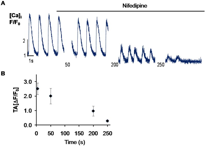
Nifedipine inhibition of Ca2+ transients. A. Representative line scan of one cell from CD117/Sca-1 and DHPR-α2 enriched cells in differentiating media after application of 40 μM nifedipine. Cells were continuously stimulated with 0.7 Hz, 80V, 5ms electrical pulse. B. Mean transient amplitude (TA, ΔF/F0) at sequential time points following application of nifedipine. Values represent Mean + SEM, n=3 cells. Supplemental Video-3 demonstrates that not all calcium transients were inhibited by the L-type calcium channel blocker.