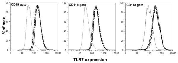
Figure 6. Level of TLR7 expression is similar between NZM2328, NOD and NZM/NOD F1 mice.
Spleen cells from 2-3 month old mice (n=3 per strain) were stained with anti-CD19 (B cells), anti-CD11c (dendritic cells), anti-CD11b (macrophages) and anti-TLR7. Figure shows TLR7 staining from representative NZM2328 (– –), NZM/NOD F1 (■ ■ ■ ■), and NOD (- - - -) mice in the CD19, CD11b and CD11c gates. The gray line represents staining with normal rabbit IgG. Similar results were obtained in an additional group of mice.

