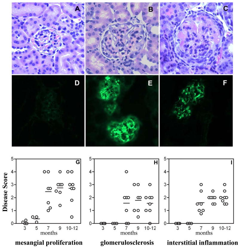
Figure 8. Characterization of renal disease in the female NZM/NOD F1.
Top panel: Representative photomicrographs of H&E-stained kidney sections of F1 mouse showing acute proliferative GN (B) and chronic GN (C). Shown for comparison is normal kidney from a NOD mouse (A). Immune-complex and C3 deposition in kidneys was studied by direct immunofluorescence staining. Representative pictures for IgG (E) and C3 (F) deposits in the F1 mice are shown. IgG deposition in NOD mice (D) was not seen. Lower Panel: Female mice were euthanized at different ages and kidney sections stained with H&E were evaluated for renal pathology. All parameters were scored on a scale of 0-5 with 0 = no disease and 5 = maximal severity. Each data point represents one mouse.

