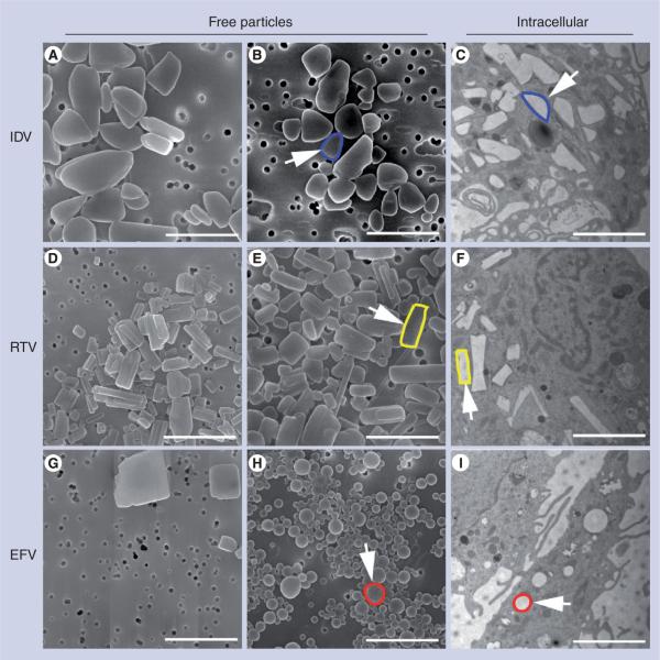
Figure 1. Nanoparticle morphology.
Scanning-electron microscopy analysis (magnification: 15,000×) of nanoformulations shown include (A) IDV-1, (B) IDV-4, (D) RTV-1, (E) RTV-4, (G) EFV-1 and (H) EFV-3 on top of a 0.2 μm polycarbonate filtration membrane. Scale bar = 2.0 μm. (A) IDV-1 and (B) IDV-4 showed ellipsoid structures with sizes of approximately 1 μm; (D) RTV-1 and (E) RTV-4 showed rod structures with sizes of approximately 550 nm; (G) EFV-1 showed cuboidal structures with sizes of approximately 600 nm while (H) EFV-3 showed spherical structures with sizes of approximately 300 nm. Transmission-electron microscopy (magnification: 15,000×) demonstrated uptake of nanoART into monocyte-derived macrophages exposed to (C) IDV-4, (F) RTV-4 and (I) EFV-3. Within the cells, each type of nanoART is readily identifiable by shape and an example has been outlined in blue for (B & C) IDV, yellow for (E & F) RTV and red for (H & I) EFV.
EFV: Efavirenz; IDV: Indinavir; RTV: Ritonavir.

