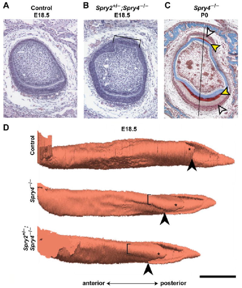
Fig. 2.

Histology and three-dimensional reconstructions of the lower incisor. Frontal histological section of (A) control and (B) Spry2+/−; Spry4−/− incisor at E18.5 (hematoxylin-eosin staining). The lingual “pouch” is indicated by the symbol ]. (C) Heidenhein's aniline blue staining of a Spry4−/− incisor at P0 shows the enamel in red and dentin and bone in blue. The red enamel (black/yellow arrowheads) is present on both labial (lower) and lingual (upper) side. Labial (lower) and lingual (upper) ameloblasts (blank arrowheads) are visible. The midline is located on the right side of the picture. The black line represents the middle axis along which measurements were made. (D) 3D reconstructions of the lower jaw incisor enamel organ at E18.5 in control, Spry4−/−, and Spry4−/−; Spry2+/− incisors. The lingual “pouch” is indicated by the symbol [. Such a pouch was not present in wild-type (control) incisors at the same stage. The lingual part of the cervical loop is indicated by an asterisk. The black arrowhead points to the split of the apical portion of the enamel organ into the lingual and labial parts of the cervical loop. Note the markedly longer lingual part of the cervical loop in sprouty mutant mice. Bar, 500 μm.
