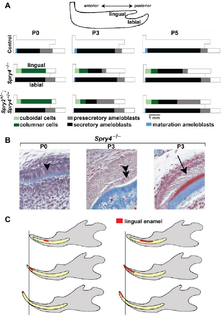
Fig. 7.

Schematic representation and frontal sections of postnatal lower incisor. (A) The relative length of the area of different types of epithelial cells on the lingual and labial surfaces is shown along the antero-posterior axes. The relative extent of the region of presecretory, secretory or maturation ameloblasts and of cuboidal or columnar cells were calculated as a proportion from the total antero-posterior length of the corresponding incisor enamel organ. The representative length of an incisor enamel organ at P0, P3 or P5 was calculated as the average of the length of the enamel organs of all the control, Spry4−/− and Spry2+/−; Spry4−/− incisor. On the lingual surface of the mutant incisor tip, cuboidal and low columnar cells are present. Compared with the labial surface, ameloblast polarization and secretion are retarded on the lingual side of mutant incisors. Note loss of presecretory ameloblasts during P3 and P5 in Spry4−/− mice. (B) Three types of epithelial cells on the lingual side of a Spry4−/− lower incisor are documented on frontal histological sections (Heidenhein's aniline blue staining): columnar cells (single arrowhead) at P0, cuboidal-low columnar cells (double arrowhead) in the anterior part of the lower incisor enamel organ at P3 and secretory ameloblasts (arrow) in the middle part of the lower incisor enamel organ at P3. The Spry4−/− incisor at P3 shows the enamel in red and dentin and bone in blue. (C) A model for how ectopic lingual enamel shifts anteriorly on the surface of the erupting incisor in Spry4−/− mice. The timing of disappearance of the lingual enamel depends on the posterior extent of the ectopic enamel zone. Variability was detected in the phenotype, such that when the length of the enamel zone was short, the enamel was abraded (disappeared) earlier (left) than in cases when the enamel zone was longer in the posterior direction (right).
