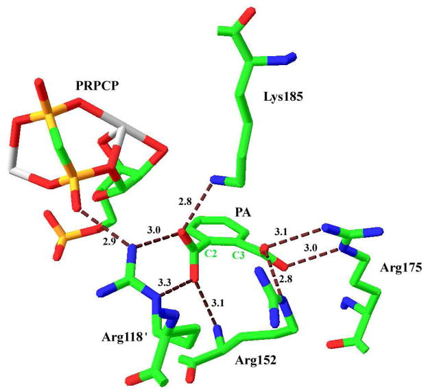
Figure 2.
Binding site for QA in the M. tuberculosis QAPRTase•PA•PRPCP•Mn2+ Michaelis complex (PDB entry 1QPR). Residues that interact with QA are shown and their hydrogen-bonding distances are represented in Å. The primes on Arg118′ designates residue from the adjacent subunit. C2 and C3 are designations of the homologous positions on QA. The Two Mn2+ are coordinated by PRPC are shown in gray. This figure was generated using SwissPDB Viewer (23).

