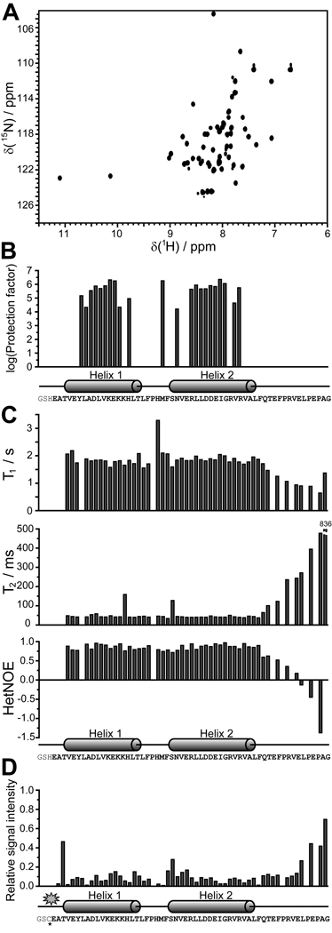
Figure 2. Identification of the structured region in the Qua1 region of GLD-1 by NMR spectroscopy.
(A) 1H,15N-HSQC spectrum for the Qua1 region. (B) The H/D exchange experiment confirms that the two predicted helices are structured and thus protected against proton exchange with the solvent. (C) 15N-relaxation results suggest that the structured core of the Qua1 region includes both helices and a short, not very flexible linker. (D) Paramagnetic relaxation enhancement of a spin-labeled Qua1 region shows strong signal attenuation toward the C-terminal end of helix 2, suggesting either an antiparallel orientation of two monomers or a helix-turn-helix monomer.

