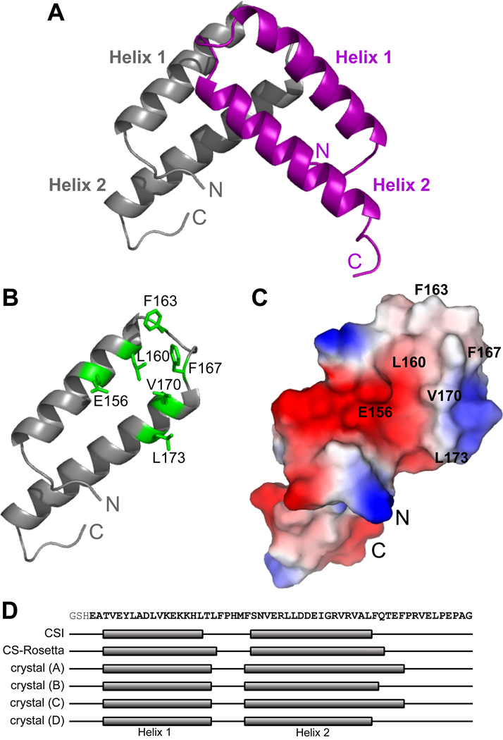
Figure 4. X-ray crystal structure of the homodimeric Qua1 region.
(A) Structure of the Qua1 homodimer. Two helix-turn-helix monomers are stacked in a perpendicular orientation. The first N-terminal and the C-terminal 5–13 residues are disordered in the electron density map. (B) The dimer interface consists of a hydrophobic patch (highlighted in green). (C) Surface charge potential of the Qua1 monomer. Dimer interface residues are labeled. (D) Mapping of the secondary structure determined from chemical shift indices (CSI), the CS-Rosetta model and all four monomers within the asymmetric unit of the crystal structure onto the Qua1 sequence.

