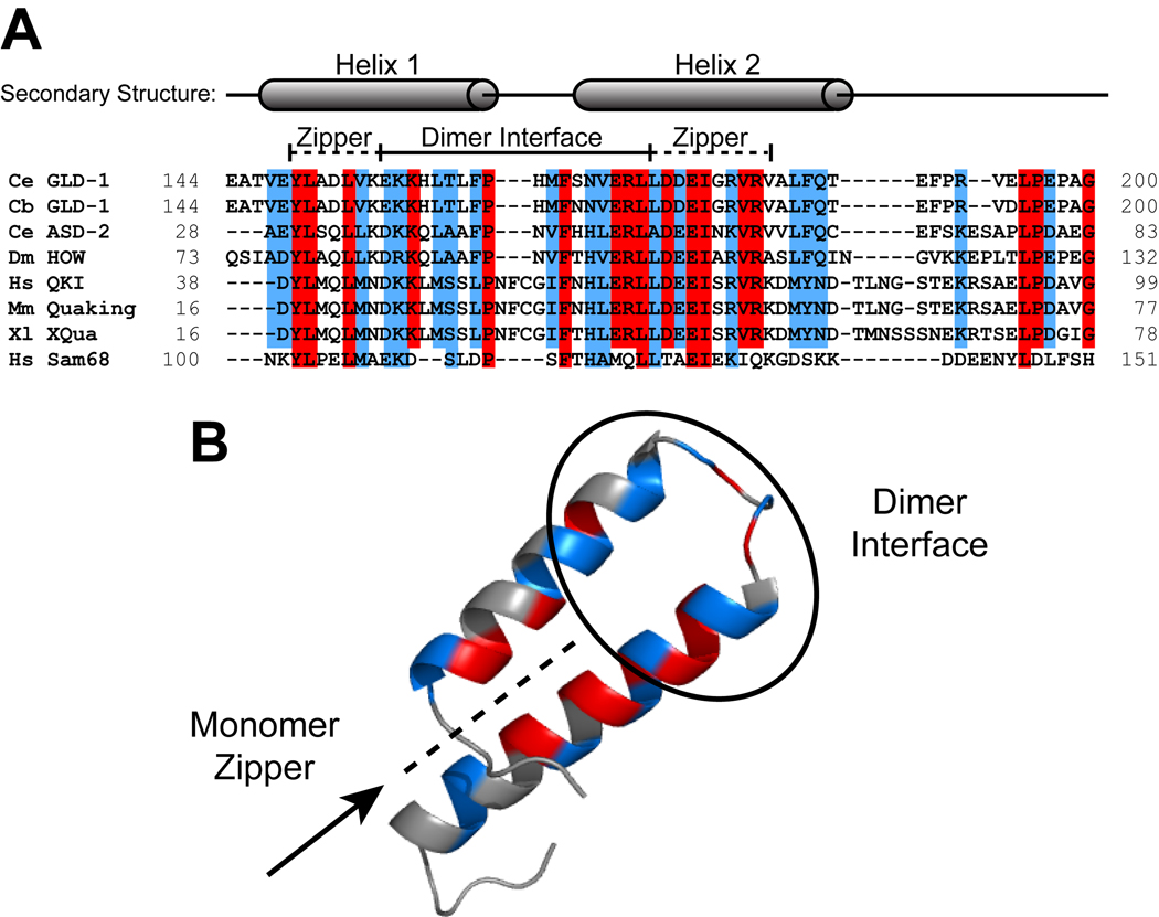
Figure 7. Sequence alignment of the Qua1 region for representative members of the STAR/GSG family.
(A) Sequence alignment. The secondary structure is mapped on top of the sequences. Identical residues are highlighted in red and similar residues in blue. The dimer interface and zipper regions are marked by a bar. (B) Identical (red) and similar (blue) residues mapped onto the GLD-1 Qua1 crystal structure.

