
Figure 2.
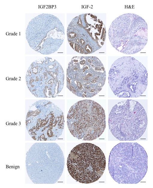
Immunohistochemical staining for IGF2BP3 and IGF-2 by tumor grade. Representative tissue microarray cores of pancreatic ductal adenocarcinomata with immunohistochemical staining for IGF2BP3 and IGF-2 by tumor grade. Note the complete lack of immunohistochemical staining for IGF2BP3 in benign pancreatic tissue. While there appears to be positive staining for IGF-2 in the benign cores, this was exclusively acinar staining and not ductal. [Scale bar, 100 μm.]