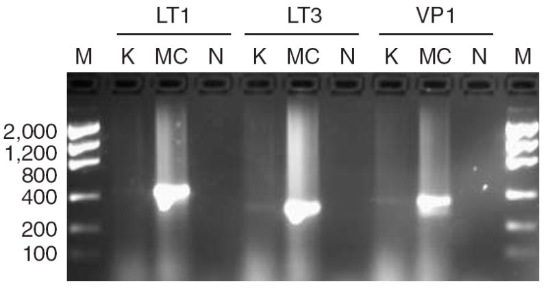
Figure 1. Detection of polyomavirus in a single keratoacanthoma.

Genomic DNA (50 ng) from a keratoacanthoma (K) or a Merkel cell carcinoma (MC) were amplified with primers specific for the LT1, LT3, or VP1 genes of polyomavirus associated with Merkel cell carcinomas (MCCs). N, non template negative control; M, low mass DNA ladder (fragment length in base pairs is indicated).
