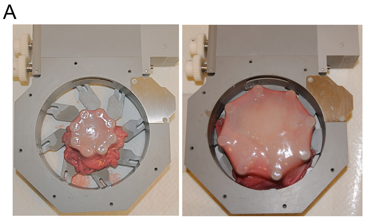
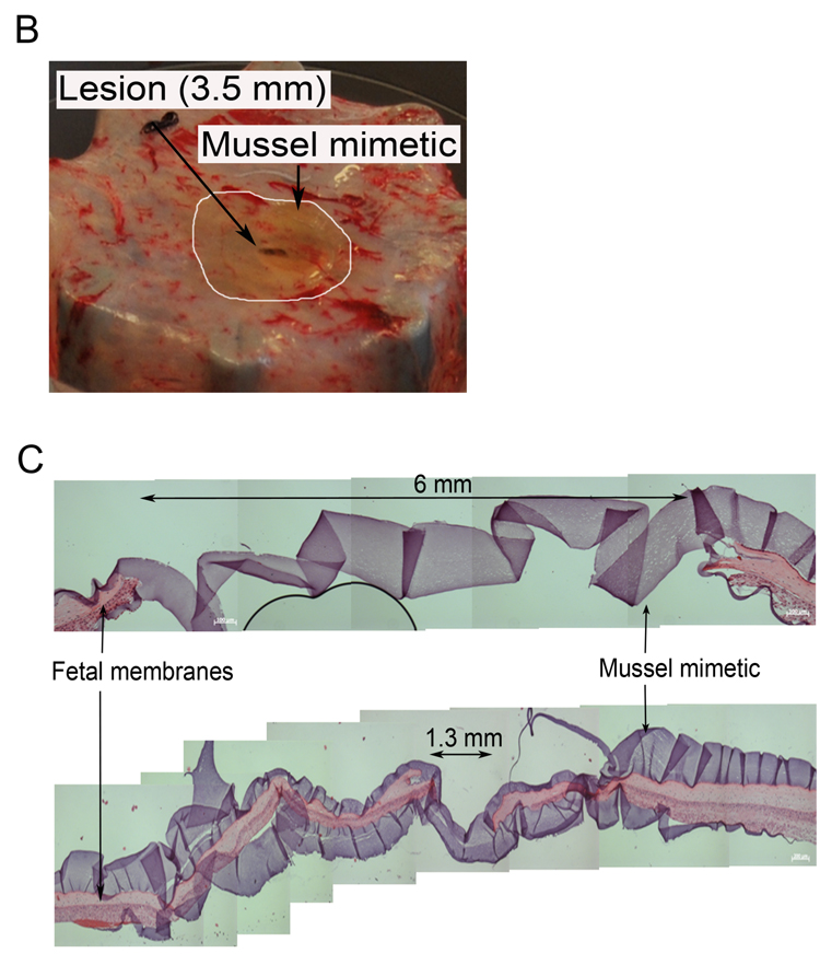
Figure 2.
Ex vivo sealing of fetal membrane defects with mussel-mimetic adhesive. (A) Fetal membranes mounted in a computerized radial stretch device before and after stretch. (B) Through-thickness puncture wounds (arrow) were created on fresh fetal membranes with a Ø 3.5mm trocar. Approximately 0.5 mL of adhesive was applied over the defect. The white line marks the area of sealant. (C) Hematoxylin/eosin stained cross-section of a trocar puncture treated with cPEG adhesive. The hydrogel appears as ribbon-like structure that bridges the puncture edges. The bottom image shows a cross-section of the same lesion at a narrow location.


