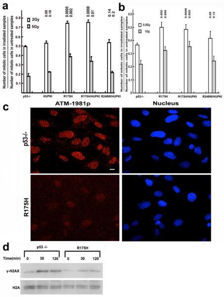
Figure 4. Impaired G2-M checkpoint and ATM activation in R175H mutant cells after irradiation.
G2-M checkpoint in MEFs (a) and proliferating thymocytes (b) of various genotypes. Mean values from three independent experiments are shown with standard deviation. P values between p53−/− and other samples are indicated. (c) IRIF of ATM-1981p in p53−/− and R175H MEFs 30 mins after 2Gy irradiation. Nucleus is countered stained with DEPI. (d) Phosphorylation of H2AX (γ-H2AX) in p53−/− and R175H mutant MEFs after 2Gy of IR.

