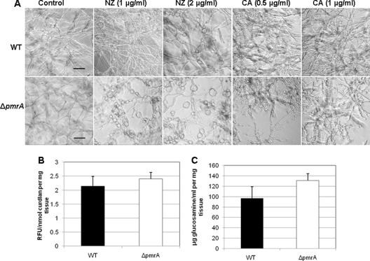
Fig. 2.
(A) Effect of cell wall inhibitors on the WT and ΔpmrA strains. A total of 103 conidia/ml of the WT and ΔpmrA strains were inoculated onto coverslips immersed in GMM medium and grown in the presence or absence of the indicated concentrations of nikkomycin Z (NZ) or caspofungin (CA) for 16 to 20 h at 37°C. Coverslips were removed and observed by microscopy. Scale bar, 20 μm. (B) β-1,3-Glucan content was quantified using the aniline blue assay as previously described (8, 25, 26), using curdlan, a β-1,3-glucan analog, as the standard. Values are expressed as relative fluorescence units (RFU) per mg of mycelial tissue. (C) Quantification of chitin was performed based on a modified protocol (8) and reported as glucosamine equivalents (μg/ml).

