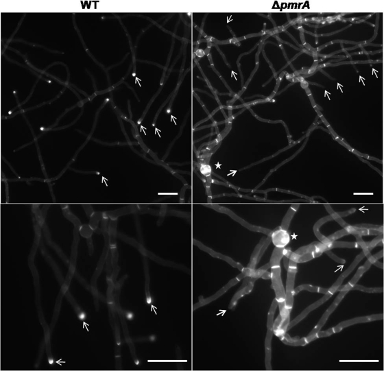
Fig. 3.
Calcofluor white staining of the WT and ΔpmrA strains. A total of 103 conidia/ml of the WT and ΔpmrA strains were inoculated onto coverslips immersed in GMM medium and grown for 24 h at 37°C. The coverslips were rinsed in sterile water and inverted onto a 200-μl drop of calcofluor white staining solution for 5 min at room temperature. Coverslips were then rinsed twice for 10 min in sterile water and mounted in Cytoseal 60 mounting medium. To ensure that the results of the calcofluor white staining of different samples were appropriately compared, the ultraviolet light exposure time for each sample was set at 25 ms for the WT untreated control, and the strains were exposed for the same lengths of time. Scale bar, 50 μm.

