Figure 1.

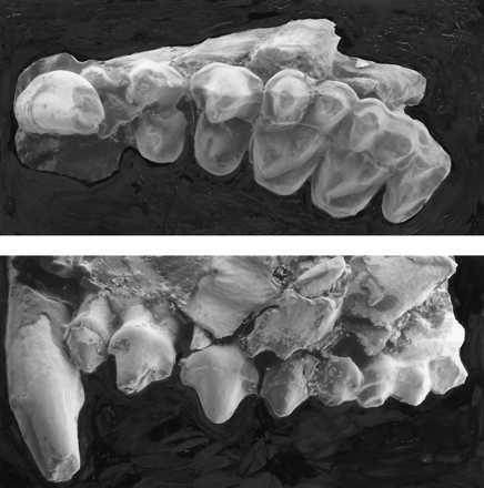
Maxillary dentition of P. sylviae DPC 15518. Scanning electron micrograph crown view (Upper) and lateral view (Lower). Magnification, ×11. Note anterior groove on canine, large P4, subequal M1–2 with distinct hypocones, and small M3.

Maxillary dentition of P. sylviae DPC 15518. Scanning electron micrograph crown view (Upper) and lateral view (Lower). Magnification, ×11. Note anterior groove on canine, large P4, subequal M1–2 with distinct hypocones, and small M3.