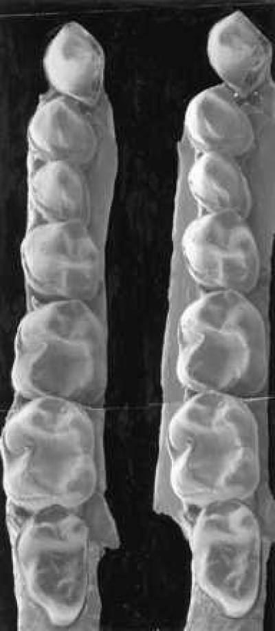
Figure 2.

Mandibular dentition of P. sylviae. Stereopair scanning electron micrograph of left lower partial dentition of CGM 42209. Magnification, ×11. Canine and M3 are reversed from a micrograph of DPC 15416, a right lower partial dentition. Note large P2, size disparity between P3 and P4, the twinned hypoconid-entoconid on M1 and M2, and the small size of M3.
