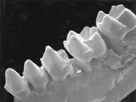
Figure 3.

Scanning electron micrograph of P. sylviae. Magnification, ×8.1. Three quaters lateral view of tooth crowns in CGM 42209. Note the height differenence between the molar trigonids and talonids, the retention of a paraconid on M1, and its loss on M2.
