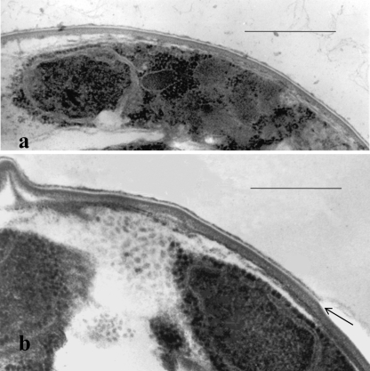
FIG. 1.
Transmission electron micrographs of thin sections of peripheral portions of glutaraldehyde-formalin-fixed C. parvum whole oocysts showing a peripheral sporozoite with a nucleus (a) and perpendicular cross sections through the oocyst wall (a and b). Note the four-layer structure of the wall and, in the outer portion of the wall, a membrane-like electron-translucent layer, which appears to be split along the axis of the translucent zone (as indicated by the arrow in panel b). Bars, 0.5 μm.

