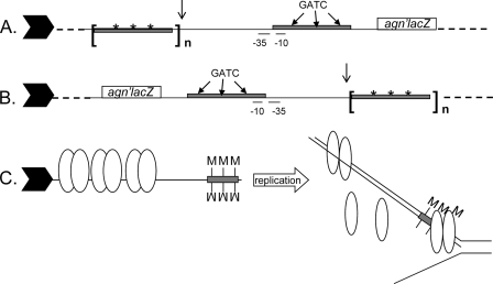
FIG. 4.
Schematic of important elements of the reporter fusion of agn′-lacZ showing the relative positions of the additional OxyR binding sites and the orientation in the chromosome (not to scale). Indicated are the promoter elements (−10 and −35) and the Dam target sequences (GATC). Gray rectangles indicate OxyR tetramer binding regions, asterisks indicate mutated GATC sequences, and n indicates the number of OxyR binding sites. The line arrow indicates the insertion position of the −112 or −466 element in different isolates (Table 3). The filled block arrow indicates the direction of the passage of the replication fork. (A) Orientation relative to the replication fork for isolates except MV1311 and MV1314. (B) In MV1311 and MV1314 the sequence elements between the dashed lines were inverted on the chromosome. (C) Schematic model depicting how three upstream auxiliary OxyR binding sites can bias phase variation to the off phase. Symbols are described above; each oval represents an OxyR dimer, M designates a methyl group, and the open arrow indicates DNA replication. Only half of the replication fork is shown.

