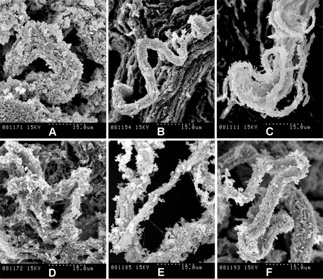
FIG. 5.
Spreading pellicles fixed with conventional methods for SEM show the organization of bacilli in the cords. Shown are SEM micrographs of cords formed in liquid medium by rough colonies of M. chubuense (A), M. gilvum (B), M. marinum (C), M. obuense (D), M. parafortuitum (E), and M. vaccae (F). The samples were processed following conventional SEM procedures of fixation with aldehydes, and the extracellular components are greatly extracted, rendering the cell surface visible. Note the differences in cord morphology among the species. The images are representative of images obtained from four different cultures.

