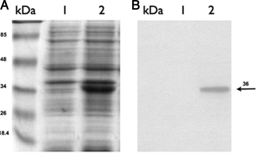
FIG. 2.
SDS-PAGE and Western blotting of S. Typhimurium strains. (A) Proteins of the membrane fraction were separated by SDS-PAGE and stained with Coomassie brilliant blue. (B) Western blot analysis using a monoclonal antibody against Synechocystis Δ12-desaturase shows that it was present in the membrane fraction of Stm(pΔ12). Lanes kDa, protein markers; lanes 1, membrane proteins from Stm(pNir) cells; lanes 2, membrane proteins from Stm(pΔ12). The arrow indicates the position of the Δ12-desaturase, with an apparent molecular mass of 36 kDa.

