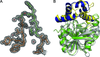
FIG. 2.
Crystal structure of Cif. (A) 2Fo-Fc map of the final refined electron density of Cif-WT, contoured to 1σ; the HGFG motif (residues 61 to 64) is colored green. A density mask with a 1.8-Å radius has been applied to prevent the display of electron density from neighboring residues. (B) Ribbon diagram depicting the superposition of ArEH and Cif-WT by DaliLite v.3 (28). The Cif core domain is colored in gray, and the cap domain is in yellow; ArEH is colored with a green core and a blue cap domain.

