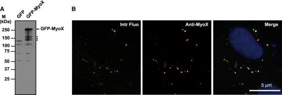
FIG. 1.
Biochemical characterization of the anti-MyoX antibody. (A) The rabbit polyclonal anti-myosin-X antibody identified a polypeptide band of the expected molecular mass in lysates of GFP-MyoX-expressing CHO cells. In contrast, no specific band was detected in lysates of GFP-expressing CHO cells, indicating that no MyoX is expressed in CHO cells. The arrow points out intact GFP-MyoX, whereas arrowheads indicate degradation products. M, molecular mass markers. (B) CHO cells expressing GFP-MyoX were immunofluorescently stained for MyoX. We observed that the intrinsic fluorescence of the GFP tag (Intr Fluo) colocalized with the immunostaining of MyoX (Anti-MyoX), thus attesting to the specificity of the antibody for MyoX.

