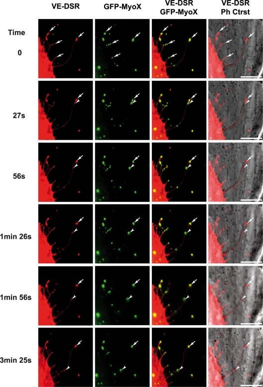
FIG. 10.
Selected sequence focusing on the coordinated backward movements of MyoX and VE-Cad. Subconfluent HUVECs, transiently cotransfected with plasmids expressing GFP-MyoX and VE-DSR, were observed by video microscopy at 17 h posttransfection at a frame rate of 1 image/3 s. Full arrowheads and arrows indicate moving and immobile patches, respectively, for VE-Cad and MyoX. Bars, 10 μm.

