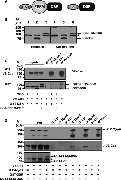
FIG. 13.
Biochemical characterization of the recombinant proteins GST-FERM-DSR and GST-DSR. (A) Schematic representation of MyoX and its derived fragments. The recombinant fragment GST-FERM-DSR was designated to keep the capacity of MyoX to interact with VE-Cad via its FERM domain. The GST-DSR recombinant fragment was used as a control. (B) Expression of GST-DSR and GST-FERM-DSR. CHO cells were transfected with either GST-DSR (lanes 2 and 5) or GST-FERM-DSR (lanes 3 and 6) or not (lanes 1 and 4). Expression of GST-DSR and GST-FERM-DSR in CHO cells was verified by Western blotting (WB) using the anti-GST antibody. This antibody recognized an 80-kDa band and a 110-kDa band corresponding to GST-DSR and GST-FERM-DSR, respectively. Additionally, it also unspecifically recognized a protein present in CHO lysates (asterisk). (C) GST-FERM-DSR interacts with VE-cadherin. Anti-VE-Cad immunoprecipitations (IP VE-Cad) were performed on CHO cell lysates coexpressing either VE-Cad and GST-FERM-DSR or VE-Cad and GST-DSR using the Mab anti-VE-Cad BV9. As controls, aliquots of whole CHO lysates (inputs) and immunoprecipitations performd on CHO lysates using mouse nonimmune IgG (IP Ctrl) were analyzed in parallel. A 110-kDa band corresponding to GST-FERM-DSR was detected in the anti-VE-Cad immunoprecipitate, indicating that GST-FERM-DSR is able to interact with VE-Cad. The anti-GST antibody recognized, as in panel B, GST-DSR and GST-FERM-DSR, as well as an unspecific protein present in CHO cell lysates (asterisk). (D) Celluar VE-Cad sequestration by the MyoX FERM domain. VE-Cad-expressing CHO cells (40) were transiently transfected with plasmids coding for GFP-MyoX and either GST-DSR or GST-FERM-DSR. After cell lysis, anti-MyoX immunoprecipitations were resolved on 4 to 12% gradient gels, electrotransferred, and probed successively for VE-Cad, MyoX, and GST. As controls, aliquots of the various CHO lysates and immunoprecipitations performed using rabbit nonimmune IgG (IP Ctr) were analyzed in parallel. Molecular markers (M) are given at the left (B to D).

