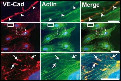
FIG. 5.
Localization of VE-Cad in subconfluent HUVECs. Images in the central row show a HUVEC monolayer immunolabeled for VE-Cad (red, left) and actin (green, center), as well as the merge (right). The selected enlargements of a mature cell-cell junction (continuous frame) and an immature junction (dotted frame) are shown in the upper and lower rows, respectively. In mature junctions, VE-Cad and cortical actin fibers are located at the cell circumference (arrowheads, upper row). In contrast, in immature junctions, VE-Cad colocalizes with radial actin fibers along filopodial extensions bridging adjacent cells (arrows, lower row). Bars: 2 μm, upper; 10 μm, middle; and 5 μm, lower.

