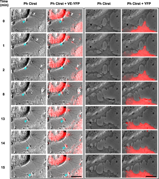
FIG. 7.
Selected sequences showing VE-YFP moving along filopodia. Subconfluent HUVECs were transiently transfected with the plasmids expressing either VE-YFP or YFP. At 17 h posttransfection time, confocal video microscopy images were taken every min for 1 h both in phase-contrast (Ph Ctrst) to see the filopodia and in the yellow fluorescence channel. Panels corresponding to VE-YFP-expressing HUVECs (left) showed two distinct patches, indicated by arrowheads, moving sequentially along two filopodia (see Movie S1 in the supplemental material). In contrast, no YFP was detected along filopodia, as illustrated in panels corresponding to YFP-expressing HUVECs (right) (see Movie S2 in the supplemental material). Bars, 10 μm.

