FIG. 4.
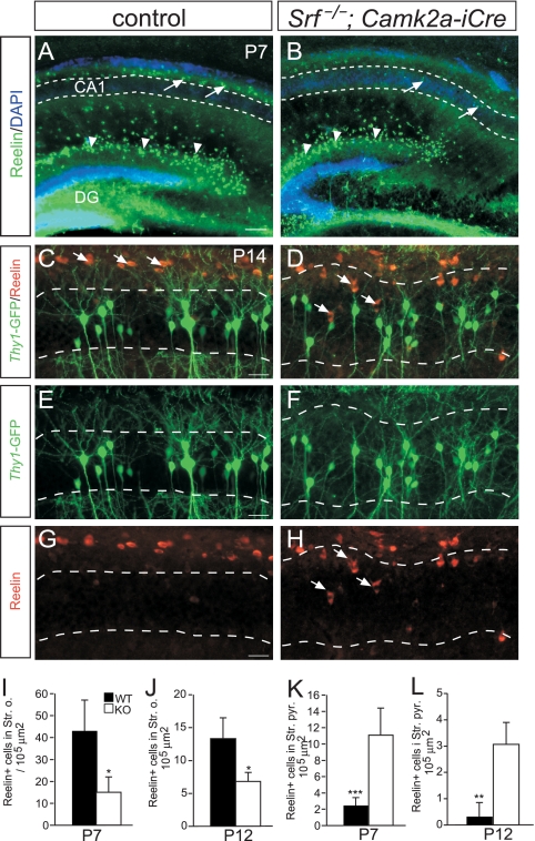
Reelin-positive cells are misplaced in Srf mutant hippocampi. (A and B) Overview of reelin distribution in the hippocampi of P7 WT (A) and Srf mutant (B) mice. In Cajal-Retzius cells of the DG (arrowheads), no obvious changes in reelin expression were noticed between the genotypes. In Srf mutants, reelin-positive cells entered the CA1 cell body region (arrows in panel B), whereas in WT mice (arrows in panel A), they remained restricted to areas outside CA1 (indicated by the dashed lines in panels A and B). (C to H) In WT P14 mice (C, E, and G), reelin-positive cells were confined to positions above the CA1 stratum pyramidale (indicated by dashed lines). In Srf mutants (D, F, and H), reelin-positive cells enter the CA1 stratum pyramidale (arrows in panels D and H). Thus, in Srf mutants, ectopic dendritic arborizations inside the stratum pyramidale are next to reelin-expressing cells. (I to L) Quantification of reelin localization in the stratum oriens (I and J) and the stratum pyramidale (K and L) at P7 (I and K) and P12 (J and L). Scale bars: A and B, 100 μm; C to H, 20 μm.

