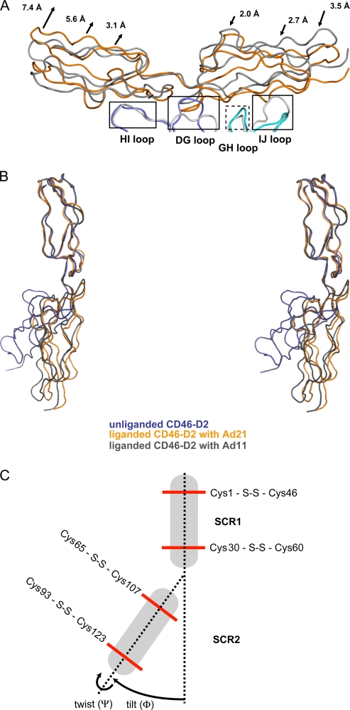
FIG. 5.
Comparison of the CD46-D2 structure in different states of ligation. (A) Superposition of the Ad21 knob-CD46-D2 complex (Ad21 knob protomers are in cyan and blue, and CD46-D2 is in orange) with the Ad11 knob-CD46-D2 complex (Ad11 knob protomers are in light gray, and CD46-D2 is in dark gray). Common binding regions are boxed. The dashed box indicates a binding region that mediates interactions only in the Ad21 knob. The arrows depict the displacement of the Ad21 knob-bound CD46-D2 regions compared to their positions in the Ad11 knob-CD46-D2 complex. (B) Superposition, based on residues in the SCR1 domain, of the three CD46-D2 structures known to date, shown in stereo. Unliganded CD46-D2 is shown in dark blue, CD46-D2 as seen in the complex with the Ad21 knob is shown in orange, and CD46-D2 as seen in the complex with the Ad11 knob is shown in dark gray. (C) Schematic view of unliganded CD46-D2. Disulfide bonds are represented with red lines. Two dashed lines link the disulfide bonds in SCR1 and SCR2. The variations in domain orientation are expressed in tilt angles (Φ) and twist angles (Ψ), as indicated with arrows (5).

