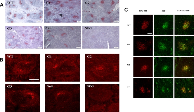
FIG. 6.
Characterization of spleen morphology in glycosylation-deficient mice. In order to assess if a lack of sugars can induce an overt phenotype in spleen, a number of morphological assays in spleen derived from transgenic, wild-type, and PrP knockout animals (null) were performed. (A) FDC networks were detected using the FDC-M2 antibody, which labels mature FDC networks within the germinal center. With light microscopy analysis using a Nikon Eclipse E800 microscope, no differences were highlighted between wild-type and glycosylation-deficient mice. (B) CD16/CD32 antibody was used to investigate the functionality of FDC by confocal microscopy analysis. Again, there were no differences evident in the labeling between the different genotypes. Scale bar, 50 μm. (C) PrP localization within the FDC network was analyzed by confocal microscopy analysis. Double immunofluorescent labeling of PrP with 1B3 antibody (green) and of FDC networks with FDC-M2 antibody (red) showed that PrPC colocalizes within the FDC networks (yellow) of each of the transgenic genotypes and wild-type samples. Scale bar, 50 μm.

