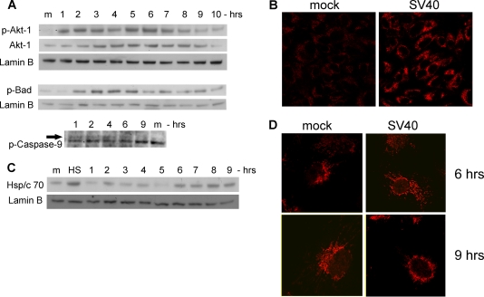
FIG. 4.
Activation of survival pathway and stress response. (A) Western blotting showing activation of Akt-1 by phosphorylation (top), upregulation of Akt-1 protein (middle), and inhibition of BAD and caspase-9 by phosphorylation. Lamin B served as a loading control. (B) Detection of phospho-Akt-1 by immunostaining. Cells were fixed with 4% formaldehyde at 6 h postinfection and stained with polyclonal anti-P-Ser473-Akt-1 and Cy3. Images were taken at a magnification of ×40. (C) Modulations of Hsp/c70 protein level. HS indicates a positive control, showing the heat shock response of CV-1 cells that were placed at 55° for 1 h. Lamin B served as a loading control. (D) Confocal microscopy of Hsp/c70. Cells were fixed at the designated time points and stained with monoclonal anti-Hsp/c70 and Cy5. Photographs were taken at a magnification of ×60 with zoom 2.

