FIG. 1.
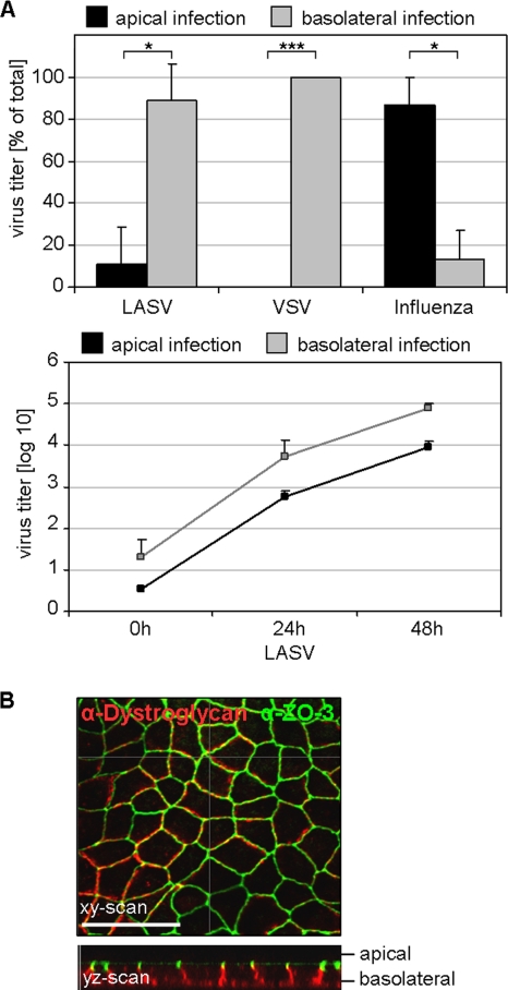
Entry of Lassa virus into polarized epithelial cells. (A) Polarized MDCK-II cells grown on Transwell filters with a pore size of 3.0 μm were infected with Lassa virus (LASV) at an MOI of 1 via either the apical (black) or basolateral (gray) route. After 48 h, total virus yield from the combined media of the lower and upper chambers was determined by TCID50 (upper panel). Influenza virus and VSV served as controls for apical and basolateral virus infection, respectively, and titers were determined at 8 h p.i. The virus titers of LASV at indicated time points are graphed in the lower panel. Average values were obtained from three independent experiments. The error bars denote standard deviations. ***, P < 0.001; *, P < 0.05 (t test). (B) Localization of the LASV receptor dystroglycan. MDCK-II cells grown to polarity on Transwell filters with a pore size of 0.4 μm were fixed with methanol-acetone and immunostained for expression of the LASV receptor dystroglycan (red) and the tight-junction-specific protein ZO-3 (green) and were subsequently analyzed by confocal microscopy. The optical section in the xy direction was scanned at the height of tight junctions. The yz scan shows distributions of dystroglycan and ZO-3 protein with respect to the apical and basolateral cell surfaces. The scale bar represents 20 μm.

