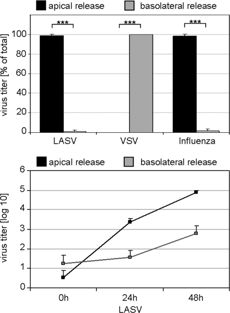
FIG. 2.
Directional release of Lassa virus from polarized epithelium. MDCK-II cells grown to polarity on 3.0-μm-pore-size Transwell filters were infected with LASV from the basolateral cell surface at an MOI of 1. Virus released into the apical chambers and into the basolateral chambers were determined by TCID50 titration at 48 h after infection (upper panel). VSV and influenza virus were inoculated from the basolateral or apical side of the cell cultures, respectively, and progeny virus was titrated 8 h p.i., as described in Materials and Methods. Additionally, LASV titers from the cell culture media of the apical or basolateral chamber were measured at the indicated time points and graphed (lower panel). Statistical evaluation was performed as described for Fig. 1.

