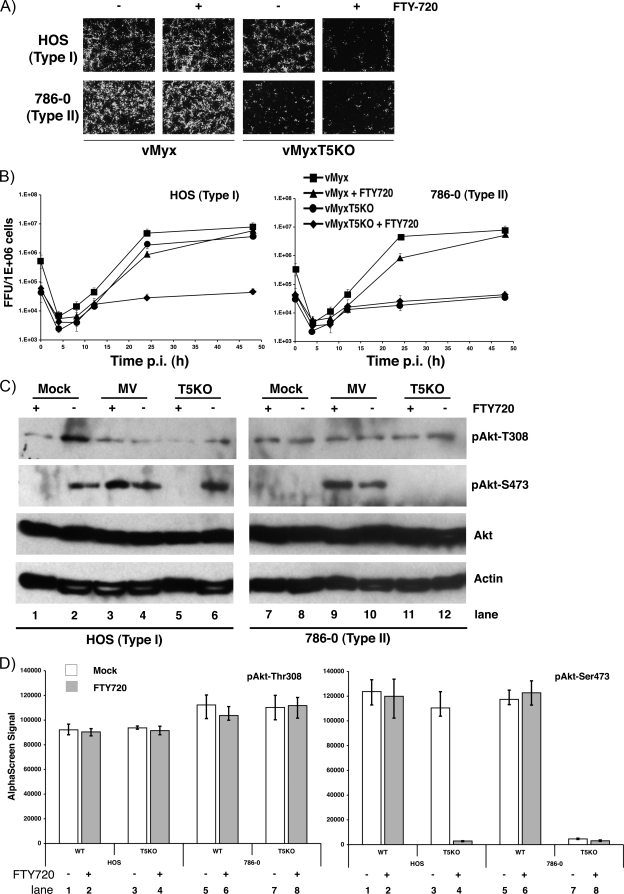
FIG. 2.
Inhibition of vMyxT5KO replication by FTY720. (A) HOS, 786-0, and SK-MEL-5 human cancer cells were infected with either vMyx-gfp or vMyxT5KO-gfp in the absence (−) or presence (+) of FTY720, and at 48 hpi, viral focus formation was determined by florescence microscopy. (B) HOS (left) and 786-0 (right) cells were infected with either vMyx-gfp or vMyxT5KO-gfp at an MOI of 1 in the in the absence or presence of FTY720, and the titer of virus collected at various hpi was determined using BGMK cells. Titers are expressed as numbers of FFU/106 cells and represent the means ± standard deviations of results from triplicate wells. (C) Representative Western blots showing detection of Akt in cell lysates from HOS (left) and 786-0 (right) cells at 24 hpi following mock infection (lanes 1 and 2), or infection with either vMyx (lanes 3 and 4) or vMyxT5KO (lanes 5 and 6) at an MOI 3 in the in the presence (+) or absence (−) of FTY720. Kinase activation of Akt is demonstrated by detection of specific phosphorylated forms pAkt-Thr308 and pAkt-Ser473. Equal sample loading was confirmed by detection of actin. (D) Cell lysates of HOS cells prepared from those shown in panel C were assayed for Akt phospho-Thr308 (left) and Akt phospho-Ser473 (right) by using the standard AlphaScreen SureFire protocol. Each sample was assayed in triplicate, and standard deviations are represented by the error bars. Mock-treated cells are represented by white bars, whereas cells treated with FTY720 are represented by gray bars. WT, wild type.

