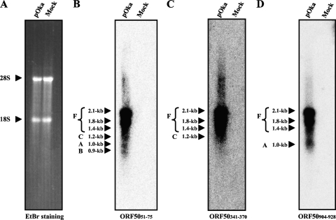
FIG. 3.
Northern blot analysis of ORF50 variants in pOka-infected MeWo cells. Total RNA was extracted from pOka-infected cells and mock-infected cells at 72 h postinfection (with full CPE in the infected cells) and separated on a formaldehyde-1% agarose gel. Ethidium bromide (EtBr) staining is shown for the size markers, and the 28S and 18S ribosomes are indicated by arrowheads (A). The blots were probed with three antisense oligonucleotides, whose positions are shown in Fig. 2B-a. The letters and size with an arrowhead or arrowheads grouped with a bracket at the left of each panel indicate the ORF50 variants predicted from each size. “F” indicates the ORF50 full-size cDNA without alternative splicing.

