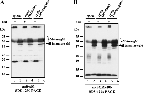
FIG. 5.
Comparison of protein expression from the ORF50 gene between rpOka, rpOkaORF50AS(−), and rpOkaORF50AS(−)Rev-infected MeWo cells. Each virus was propagated by cell-to-cell spread at a ratio of 1 infected cell to 10 uninfected cells, lysed when full CPE was observed (almost 72 hpi), and subjected to WB with anti-gM Ab (A) and anti-ORF50N Ab (B). All cells were lysed in SDS-PAGE sample buffer and either boiled (lanes 1, 3, and 5 of panels A and B) or prepared without boiling (lanes 2, 4, and 6 of panels A and B) to visualize all of the proteins translated from the ORF50 gene.

