FIG. 8.
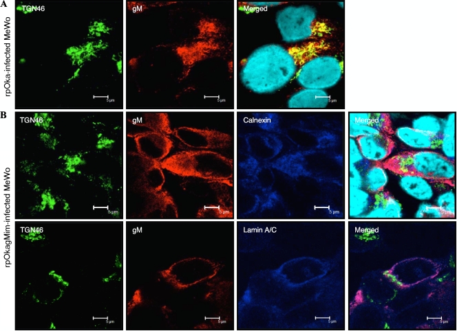
Localization of mutant gM in rpOkagMim-infected cells. MeWo cells were infected with rpOka (A) or rpOkagMim (B) cell-free virus at a multiplicity of infection (MOI) of 0.05 and fixed in ice-cold acetone-methanol at 48 hpi. The fixed cells were doubly (A) labeled for the TGN marker TGN46 (green) and gM (red) or triply (B) labeled for TGN46 (green), gM (red), and the ER marker calnexin (blue) (upper panel in B) or the nuclear membrane marker lamin A/C (blue) (lower panel in B). Nuclei were stained with Hoechst 33342 (cyan). Scale bars = 5 μm.

