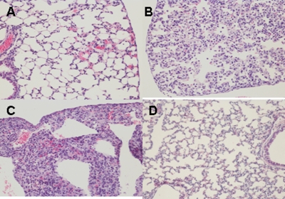
FIG. 8.
Photomicrographs of hematoxylin- and eosin-stained lung sections of mice challenged with clade 2.1 H5N1 virus at 6 days postchallenge. (A) Mice vaccinated with live BacHA; (B) mice vaccinated with inactivated BacHA; (C) unvaccinated mice challenged with virus; (D) normal morphology seen in uninfected mice.

