
Figure 1.
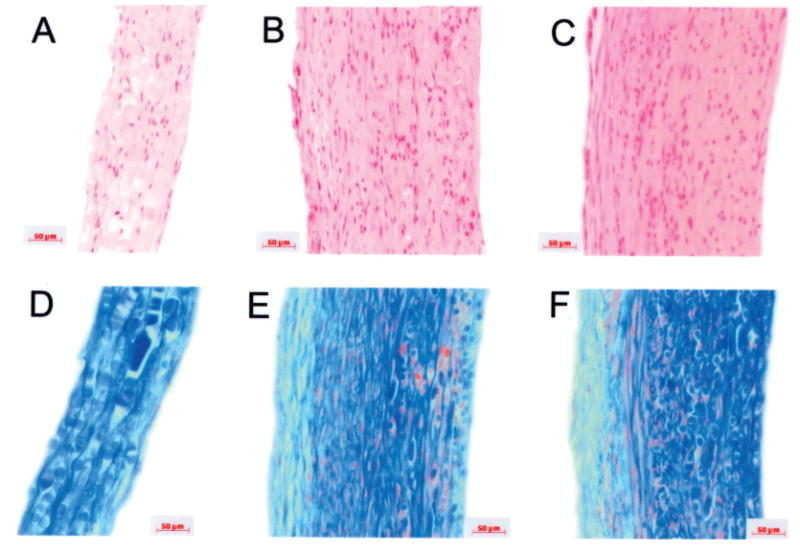
Histologies of engineered vessels. Vessels were grown at three radial distension rates: 0 beats per minute (bpm; nonstretched control), 90 bpm, and 165 bpm. (A, D) Nonstretched control vessels; (B, E) vessels cultured at 90 bpm; (C, F) vessels cultured at 165 bpm. (A–C) H&E stains; (D–F) Masson's trichrome, where collagen stains blue, cell bodies stain red, and glycosaminoglycans stain green. PGA polymer fragments more noticeable as oval or rectangular objects (D–F), staining blue. Wall thickness is increased in pulsed vessels. Scale bars: 50 μm.