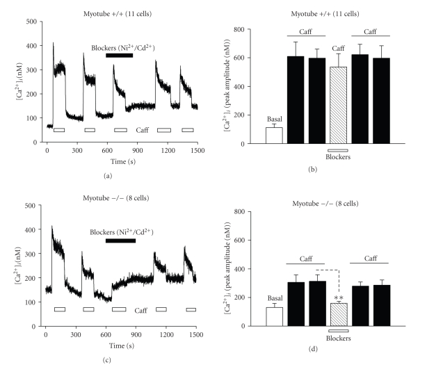
Figure 6.
Effect of caffeine on [Ca2+]i in isolated myotubes in the presence of Ca2+ channel blockers (Ni2+/Cd2+). Representative traces show the typical time course of the response to 20 mM caffeine observed in (a) wild type (+/+) myotube; (c) calpain 3-deficient (−/−) myotube, each treated with Ni2+/Cd2+. The duration of exposure to caffeine (open bars), or 50 μM Ni2+/100 μM Cd2+ (closed bars) is represented. Bar diagrams (b) and (d) summarize the peak amplitude of the [Ca2+]i response of myotubes to caffeine and CPA. The responses from the wild type myotubes (+/+; b) and calpain 3-deficient myotubes (−/−; d), are shown. Fifty μM Ni2+/100 μM Cd2+ was added to the extracellular medium prior to the second application. The number of cells tested is given in brackets.

