
Figure 2.
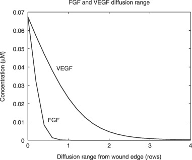
Diffusion ranges of injury‐released VEGF and FGF. Diffusion Ranges are predicted by the mathematical model and are plotted as number of cell rows from the wound edge at 60 s after injury. FGF, fibroblast growth factor; VEGF, vascular endothelial growth factors.