
Figure 6.
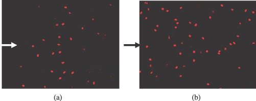
Proliferation assay using BrdU staining of HUVECs transfected with (a) empty plasmid and (b) PLCδ‐containing plasmid. Proliferating HUVECs are stained bright red. Arrows indicate the location of the wound edge on the left side. HUVEC, human umbilical vein endothelial cells.