
Fig. 7.
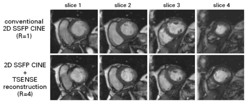
Short axis CINE images of the heart derived from unaccelerated conventional 2D SSFP imaging covering a single slice per breath-hold (top) and four-fold accelerated TSENSE 2D SSFP encompassing four slices per breath-hold (bottom). The latter supports dual breath-hold, wholeheart coverage acquisitions. The accelerated approach yielded examination time reductions of approximately 80% at 1.5 T and 3.0 T [65]. End-systolic volume and ejection fraction showed excellent correlation with the unaccelerated approach while the end-diastolic volume assessment revealed a minor difference of delta EDV =(4.1±5.8 ml) between TSENSE accelerated 2D CINE and conventional 2D CINE. (Images courtesy of Bernd J. Wintersperger, University of Munich Hospitals, Munich, Germany)