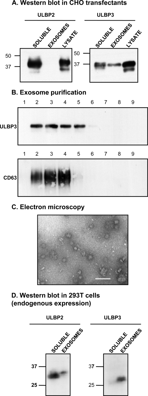
FIGURE 3.
ULBP3 is released in exosomes both in transfectants and the tumor cell line 293T. A, the exosome fraction and soluble proteins from CHO-ULBP2 and -3 transfectants were purified after 24 h in culture as described under “Experimental Procedures” and compared with total lysate from the same cells in Western blot analysis. Similar results were obtained on analysis of CV1-ULBP3 cells (supplemental Fig. 1). ULBP2 was shed mainly as a soluble protein, whereas ULBP3 was released both as a soluble protein and in exosomes. B, fractionation of exosomes in a sucrose gradient shows co-migration with CD63. C, exosomes from CHO-ULBP3 cells were negatively stained with 2% phosphotungstic acid and analyzed by electron microscopy. As expected, nano-sized vesicles from 30 to 120 nm were observed. Bar, 100 nm. D, analysis of tissue culture supernatant from the 293T cell line, which expresses ULBP2 and ULBP3 endogenously, confirmed the results obtained in the transfectant system. Soluble and exosome fractions were analyzed by Western blot. ULBP2 is mainly shed as a soluble protein, whereas ULBP3 is mainly released in exosomes.

