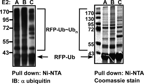
FIGURE 3.
Polyubiquitination of RFP-Ub proceeds in an E3-independent manner. RFP-Ub was absorbed onto Ni2+-NTA-agarose beads, and the beads were then added to the polyubiquitination reactions with each of the E2 enzymes. Left, Western blotting analysis with anti-RFP antibody of representative reactions. Right, similar scaled up reactions stained with Coomassie Blue. RFP-Ub ubiquitination reactions by all other E2s are documented in supplemental Fig. S7. IB, immunoblot.

