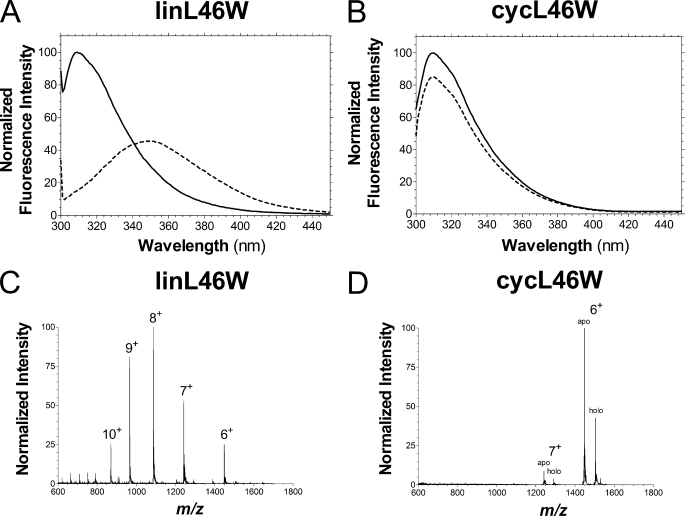
FIGURE 5.
Biophysical characterization of cycL46W and linL46W. Tryptophan fluorescence spectra (λex = 296 nm) of linL46W (A) and cycL46W (B) were obtained in the absence (dashed line) and presence (solid line) of 10 mm Mg2+. Positive mode nano-ionspray MS spectra of intact linL46W (C) and a mixture of apo- and holo-cycL46W (D) proteins were recorded as described in the text. The charge of relevant peaks are indicated.

