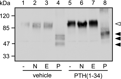
FIGURE 3.
Glycosylation does not contribute to the PTH-induced changes in molecular mass of the PTHR. CHO cells stably expressing HA-PTHR were treated with 100 nm PTH(1–34) for 12 h as indicated. The cells were lysed, and PTHR was precipitated using anti-HA affinity beads. Precipitated proteins were incubated with 50 units/ml of neuraminidase (N), 250 units/ml of Endo H (E), or 5 units/ml of PNGase F (P) for 16 h. Enzyme reactions were stopped by the addition of SDS sample buffer, and proteins were separated by SDS-PAGE. HA-PTHR was detected in Western blot analysis. The empty arrowhead indicates a fully glycosylated receptor form. The closed arrowheads indicate the different receptor forms after PNGase treatment. The positions of molecular weight standards are marked on the left.

