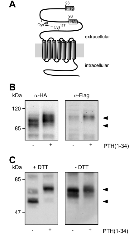
FIGURE 5.
Modification of the PTHR is restricted to the extracellular domain. A, schematic representation of the PTHR. A FLAG epitope was inserted after the signal sequence (amino acids 22 and 23), and an HA epitope was inserted replacing amino acids 93–101. A disulfide bridge between Cys48 and Cys117 stabilizing the extracellular domain is depicted by a dotted line. B, CHO cells stably expressing the FLAG-HA-PTHR were stimulated with PTH(1–34) for 12 h. The cells were lysed, and the samples were subjected to SDS-PAGE and subsequent Western blot analysis. PTHR was detected using an anti-HA antibody (left panel) or an anti-FLAG-M2 antibody (right panel). C, CHO cells stably expressing the HA-PTHR were stimulated with PTH(1–34) for 12 h. To gain better resolution of the proteins, the cells were treated 12 h prior to and throughout the experiment with 5 μg/ml tunicamycin to block N-glycosylation. The cells were lysed in SDS-buffer in the presence (left panel) or absence (right panel) of 100 mm dithiothreitol (DTT). The lysates were subjected to SDS-PAGE and subsequent Western blot analysis. PTHR was detected using an anti-HA antibody. The PTHR forms are indicated by arrowheads.

